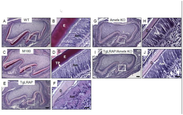
Figure 2.

Hematoxylin and eosin (H&E) staining of 5 day post natal (P5) molars from various amelogenin transgenic mouse models. (A and B) Secretory ameloblasts on distal cusp slope in WT molar (a boxed area in A) showed the typical polarized appearance of the long cylindrical ameloblast cells with Tomes’ process extensions which have laid down enamel matrix. (C and D) M180 secretory ameloblasts appeared morphologically similar to those of WT. Tomes’ process extensions were noted throughout the secretory stage. (E and F) TgLRAP secretory stage ameloblasts showed a disorganized pattern of the ameloblasts with multiple layers of cells and less elongated nuclei. Only a small amount of enamel matrix formed, and surface of dentin had an irregular pattern. (G and H) Amelogenin null molar had very thin enamel like matrix (*) and normal appearing elongated ameloblasts, but lacked Tomes’ process extensions. (I and J) Ameloblasts of LRAP transgene overexpression in amelogenin null background did not show morphological differences similar to those in amelogenin null mice. Am, ameloblast; D, dentin; E, enamel; TP, Tomes’ process. Scale bar 100 μm for A,C,E; 20 μm for B,D,F; 100 μm for G,I; 20μm for H,J.
