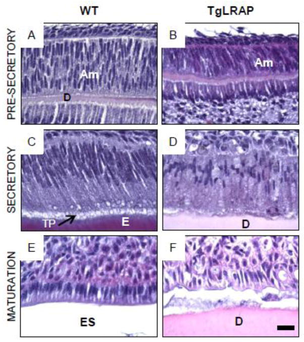
Figure 3. Stage specific phenotype of WT and TgLRAP ameloblasts.
(A and B) At the pre-secretory stage, both WT (A) and TgLRAP (B) ameloblasts were not polarized and appeared similar. At this stage enamel matrix had yet to be deposited and only a thin layer of dentin matrix was present. (C and D) At the secretory stage the WT ameloblasts (C) had elongated and were well polarized with Tomes’ process facing the enamel matrix at their apical end. In contrast, the TgLRAP ameloblasts layer (D) was less organized and only a minimal amount of enamel matrix was secreted. Tomes’ processes were not detectable in the TgLRAP cells. (E and F) In WT mice maturation stage ameloblasts (E) had transitioned to a shortened cell that was still well polarized with a widened enamel space left by the demineralized enamel matrix. The TgLRAP ameloblasts (F) had also transitioned to a shortened cell phenotype, but remained disorganized appearance. The lack of an enamel space suggests that only a thin layer of enamel was formed. Am, ameloblast; E, enamel; ES, enamel space; D, dentin; TP, Tomes’ process. Scale bar 20 μm.

