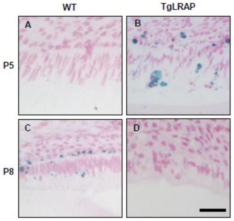
Figure 7. TUNEL staining on P5 and P8 molar ameloblasts.

(A) P5 WT molars had no TUNEL positive ameloblasts. (B) P5 TgLRAP molar showed TUNEL positive staining (in blue) in the region corresponding to the most differentiated secretory stage ameloblasts (C) AT P8 WT molars had post secretory stage ameloblasts where TUNEL positive staining was observed. (D) Ameloblasts of P8 TgLRAP mice showed post secretory morphology and slight TUNEL positive staining was observed. Scale bar 30μm.
