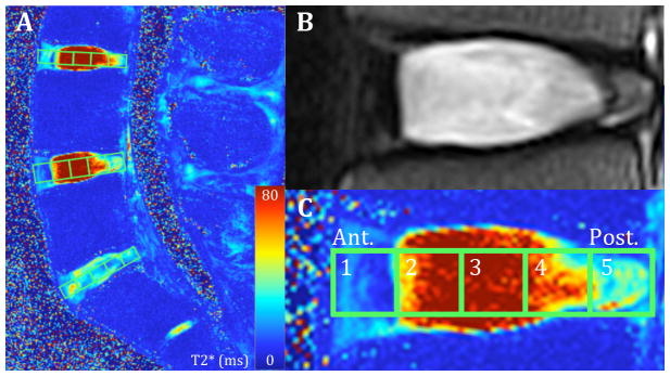
Figure 1. Magnetic Resonance Images of Lumbar Spine and Intervertebral Disc Using T2 and Quantitative T2* Techniques.

Left: T2* Map of Lumbar Spine. Top Right: Healthy Lumbar Intervertebral disc imaged with classical T2 MRI. Bottom Right: Same disc using T2* map showcasing the five regions of interest from anterior to posterior (1 to 5).
