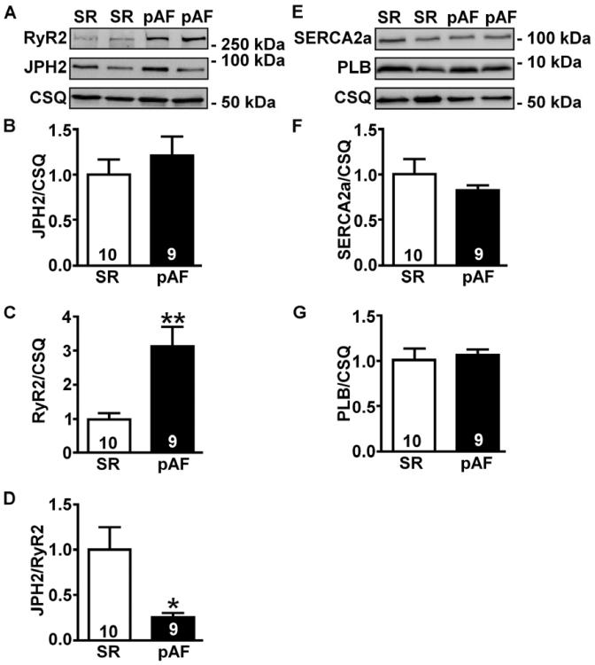
Figure 6. Reduced JPH2:RyR2 ratios in patients with paroxysmal AF.
Representative Western blots (A) and bar graphs (B–C) showing RyR2 and JPH2 protein levels normalized to calsequestrin (CSQ) in atria from patients in sinus rhythm (SR) and paroxysmal AF (pAF). (D) Ratio of JPH2-to-RyR2 levels in human atria. Representative Western blots (E) and bar graphs (F–G) showing SERCA2a and PLB levels normalized to CSQ.

