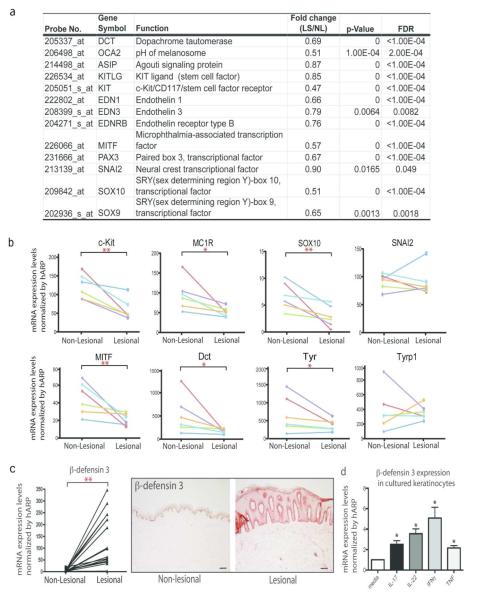
Figure 3. A broad inhibition of pigmentation genes in lesional psoriasis skin.
(a) Decreased expressions of pigmentation genes (paired lesional vs. non-lesional skin) in a meta-analysis derived of transcriptome of over 190 psoriasis patients (P<0.05, FDR<0.05) (b) qRT-PCR analysis confirms suppression of pigmentation genes in paired psoriasis lesional vs. non-lesional skin (n=6). (*P<0.05, *P<0.01). Gene expression changes for each patient were represented by a line with a different color. (c) Increased expression of β-defensin 3 in lesional psoriasis skin, compared to non-lesional skin (n=10). Bar=100μm (d) IL-17 and TNF induces the expression of β-defensin 3, an antagonist for melanocortin-1 receptor, in keratinocytes after 24 h treatment with individual cytokines: IL-17 (200ng/mL), IL-22 (200ng/mL), IFNγ (20ng/mL), and TNF (10ng/mL) (*P < 0.05; **P < 0.01; ***P<0.001, vs. Ctrl).

