
Fig. 1.
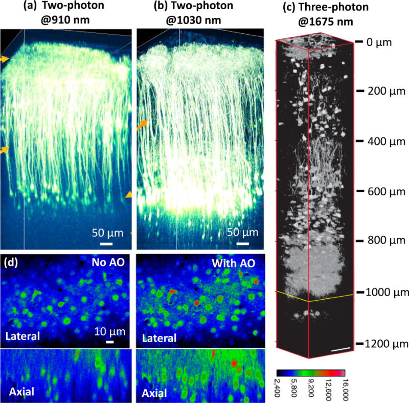
(a–c) Deep in vivo fluorescence imaging with NIR excitation. Two-photon fluorescence imaging of cortical pyramidal neurons with (a) 910-nm and (b) 1030-nm excitation in an adult eYFP-labelled mouse brain. (c) Three-photon fluorescence imaging with 1675-nm excitation of RFP-labelled pyramidal neurons in a mouse brain. (d) AO correction improves calcium imaging. Left: OGB-1 AM labeled neurons 155 μm below the brain surface without AO correction. Right: The same neurons with AO correction. Panels a-b are reprinted from R. Kawakami, et al. [1] with permission from Science; panel c is adapted from N. G. Horton, et al. [3] with permission from Nature; panel d is adapted from N. Ji, et al. [6] with permission from PNAS.