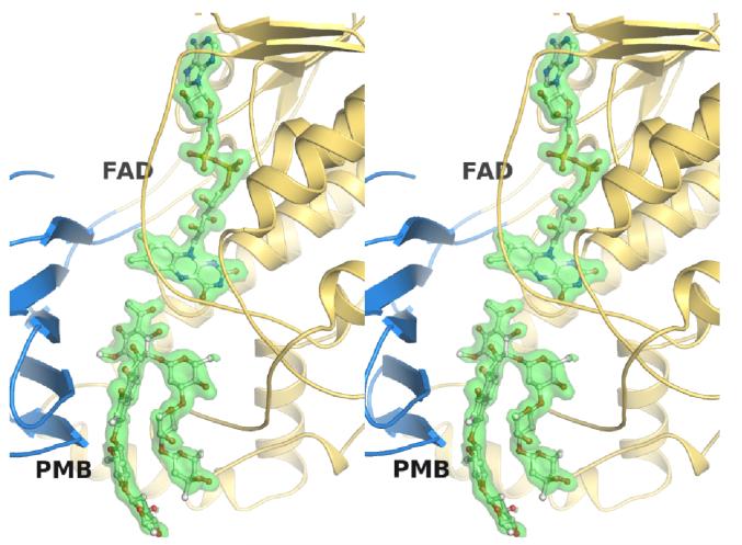
Figure 3. Wall-eye stereoview showing the electron density for FAD and premitramycin B.
The middle domain in shown in blue, the FAD domain in gold, both FAD and premithramycin B (PMB) are shown in ball and stick representation, and the electron density (SA-omit Fo-Fc map contoured to 2.5 σ) is shown as a green transparent isosurface.

