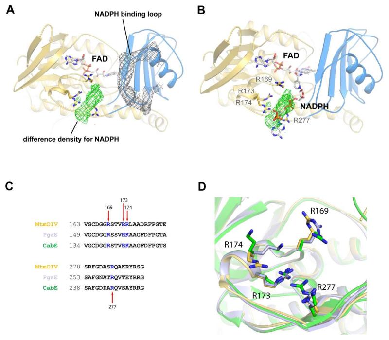
Figure 6. The low resolution crystal structure of NADPH bound to MtmOIV.
A. The ordered NADPH binding loop of MtmOIV showing 2Fo-Fc electron density (gray) contoured at 0.8 σ. Difference density (green) contoured at 2.5 σ for NADPH that was observed only in this crystal structure and not within any of the previous MtmOIV structures. The space group for the NADPH-bound co-crystal structure was P1 with 6 molecules in the asymmetric unit. The resolution was 3.5 Å and final R/Rfree values are 0.22/0.26. B. Rigid body placement of NADPH along the difference density within the MtmOIV structure which was used as a starting point for subsequent docking studies. C. Sequence alignment showing the conservation of basic residues at the putative NADPH binding site. D. Structural alignment of MtmOIV (gold), PgaE (light purple), and CabE (green) depicting residues proposed to interact with NADPH shown in stick representation.

