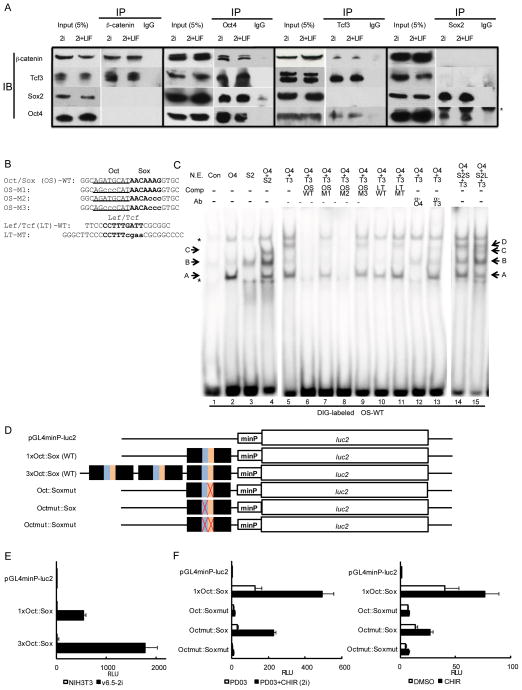
Figure 6. Physical association of β-catenin, Oct4, Sox2, and Tcf3 and in vitro binding properties of Oct4, Sox2, and Tcf3 to the Oct-Sox composite motif.
(A) Co-immunoprecipitation analysis of β-catenin, Tcf3, Oct4, and Sox2 complexes in mESCs. IP, immunoprecipitation; IB, immunoblotting. Asterisk indicates heavy chains of antibody used in IP.
(B) Sequence of oligonucleotide probes used in EMSA. Oct motif is underlined; Sox and Lef/Tcf motifs are bolded. Mutations are shown in lowercases.
(C) Cooperative bindings of Oct4, Sox2, and Tcf3 to the Oct-Sox (OS) composite motif, determined by EMSA. The indicated combinations of nuclear extracts isolated from 293T cells overexpressing Pou5f1 (Oct4), Sox2, or Tcf7l1 (Tcf3) were analyzed by EMSA using DIG-labeled OS probes. Bands A, B, C, and D denoted with arrows indicate Oct4-binary, Sox2-binary, Oct4-Sox2-ternary, and Oct4-Tcf3-ternary complexes with the Oct/Sox probe, respectively. Asterisks indicate non-specific bands. NE, nuclear extracts; Con, extracts from mock-transfected cells; O4, extracts from Oct4-overexpressing cells; S2, extracts from Sox2-overexpressing cells; S2S, smaller amount of extracts (0.2 μg) from Sox2-overexpressing cells; S2L, larger amount of extracts (2 μg) from Sox2-overexpressing cells. T3, extracts from Tcf3-overexpressing cells; Comp, unlabeled competitors; Ab, antibodies; α-O4, anti-Oct4 antibody; α-T3, anti-Tcf3 antibody. Data here is extracted from Figure S7B which provides more extensive competitor experiments and antibody supershift experiments to identity each shifted band.
(D) Schematic of luciferase reporter constructs used in (E) and (F). Pou5f1 distal enhancer region containing the Oct-Sox composite motif drives the luciferase gene with a minimal TATA-box promoter element under pGL4 vector backbone. Each mutation corresponds to mutant motifs in EMSA analysis.
(E) Luciferase reporter assay using Pou5f1 distal enhancer region in NIH3T3 cells and 2i-cultured mESCs (v6.5). Forty-eight hours after transfection with reporter constructs, cells were subjected to the assay. mESCs were maintained under 2i+LIF condition for 11 passages prior to the assay.
(F) Luciferase reporter assay in mESCs (v6.5) cultured under 2i condition (left) or CM (right). mESCs were maintained under 2i+LIF condition for 11 passages prior to the assay. Upon transfection with reporter constructs, cells were switched into basal media of 2i culture (mixture of neurobasal media, DMEM/F12, N2, and B27 supplements) with PD03 or 2i (left), or CM in the presence or absence of CHIR (right). The assay was performed 48 hours after transfection. RLU, relative light unit.

