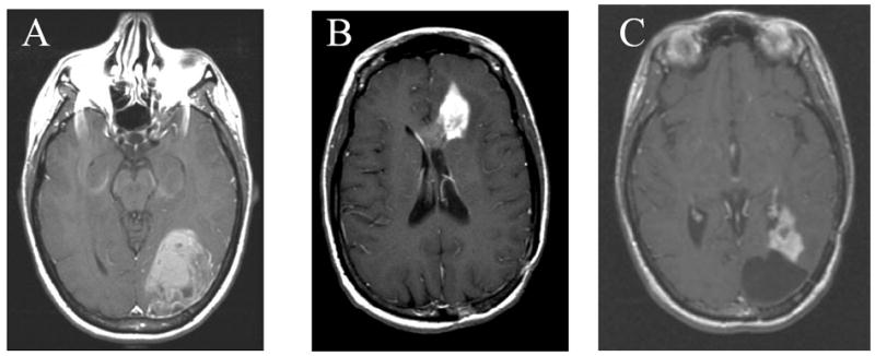
Figure 2. Post-gadolinium T1-weighted MRI of 31 year-old female approximately 20 years after whole-brain radiation and intrathecal methotrexate for acute myelogenous leukemia.

Pt had a 6 cm enhancing mass in the left occipital lobe. Pathology revealed WHO grade IV astrocytoma (GBM). (B) 14 months after her initial GBM treatment, the patient presented with a recurrence in her left frontal lobe. (C) 13 months after her GBM the patient had recurrence adjacent to her two resection cavities in the left occipital (shown) and left frontal lobes. The patient passed away 34 months after her GBM diagnosis.
