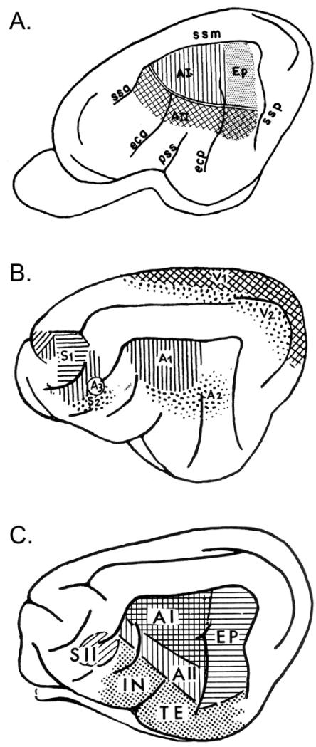
Figure 10.
Cat cortex: quick orientation and terminology. A. The basic sensory-responsive cortices from Bremer (1952). ‘A3’ is a small auditory-responsive zone within S2 (multimodal). B. Core (AI) and belt (AII, Ep) regions from Rose and Woolsey (1949), using anatomy and evoked-potential mapping (‘Ep’ = posterior ectosylvian area; ‘ss’ = suparasylvian sulcus). C. Summary of auditory-responsive regions from Ades (1959) including core (AI), belt (AII, EP), and parabelt/association (‘IN’ = insular region; ‘TE’ = temporal area) regions. Note that these regions, based on ECoG evoked-potentials, are expanded (spatially blurred) compared to current maps based on single- or multi-unit mapping.

