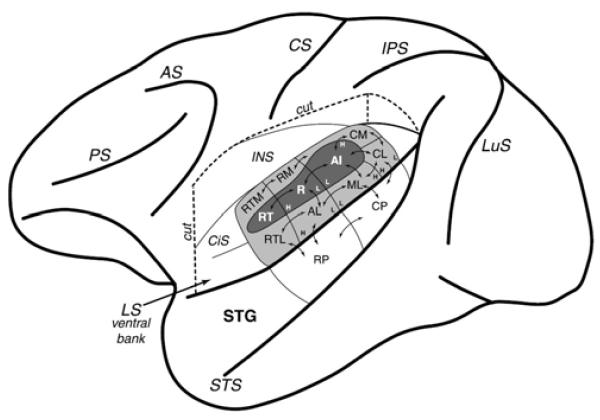
Figure 11.
Macaque cortex: core, belt, and parabelt regions from Hackett et al. (2001) (who also studied humans). ‘Core’ regions (AI, R, RT) are in dark gray within the lateral sulcus (LS, e.g. the ‘Sylvian fissure’). ‘Belt’ regions (CM, RM, RTM, RTL, AL, ML, CL) are in light gray. ‘Parabelt’ regions (RP, CP) occupy the major exposed surface of the superior temporal gyrus (STG). Note that human anatomical organization is suggested to be similar (Hackett et al., 2001, Sweet et al., 2005, Fullerton and Pandya, 2007, Brugge et al., 2008, Baumann et al., 2013).

