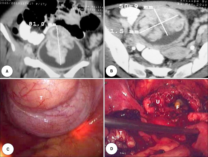
FIG. 1.
Coronal (A) and cross-sectional (B) view of the contrast-enhanced computerized tomographic scan of the pelvis showing the bladder mass. (C) Laparoscopic view of the urinary bladder with the tumor mass (T). The tumor outline was defined by cystoscopic-guided transillumination. (D) Laparoscopic view of the urinary bladder (U) after resection of the tumor mass (T). The tip of the Foley catheter (F) can be seen inside the bladder.

