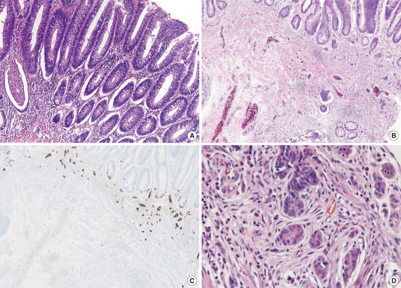
Fig. 2.
(A) A well-defined tubular adenomatous polyp is observed. The adenomatous polyp shows nuclear elongation and stratification with morphologic changes consistent with low grade dysplasia. (B) Beneath the adenomatous polyp, multiple small cellular nests are infiltrating the lamina propria and muscularis mucosae. (C) Immunohistochemical staining of chromogranin shows diffuse strong positivity in the small nests, confirming the neuroendocrine origin of the cell nests. (D) Higher magnification of the cellular nests, some fused together, composed of monotonous cells with eosinophilic and finely granular cytoplasm and central nuclei with stippled chromatin.

