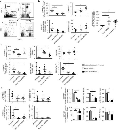
Figure 3.
Both untreated BMDC- and Dexa BMDC–administration result in a reduction in percentage and absolute number of graft infiltrating cells and an increased ratio of intragraft FoxP3+–expressing cells. (a) Gating strategy for corneal cell–infiltrating analysis. (b) The corneal infiltrating population of activated T cells (CD4+CD25+) and regulatory T cells (CD4+FoxP3+) were analyzed looking at percentage cell population, total cell number. The intragraft ratio of regulatory CD4+FoxP3+ T cells to activated CD4+CD25+ T cells was also analyzed (mean ± SEM *P ≤ 0.05 two-tailed Mann–Whitney test n = 4 per group). (c) Infiltrating population of APCs (CD11b/c+), DCs (CD11b/c+MHCII+CD86hi), and B cells (CD45RA) were evaluated, as were (d) activated NKT (CD3+CD8+CD161++), NK (CD3−CD8+CD161++) (mean ± SEM *P ≤ 0.05 two-tailed Mann–Whitney test n = 4 per group). (e) mRNA analysis of intragraft cytokine expression (normalized to β-actin, fold change relative to untreated allogeneic Tx controls) for proinflammatory cytokines IL-6, IFN-γ, and IL-1β and IDO, PD-L1, and IL-10 expression (mean ± SEM *P ≤ 0.05 two-tailed Mann–Whitney test n = 4 per group). BMDC, bone marrow–derived dendritic cell.

