Figure 3.
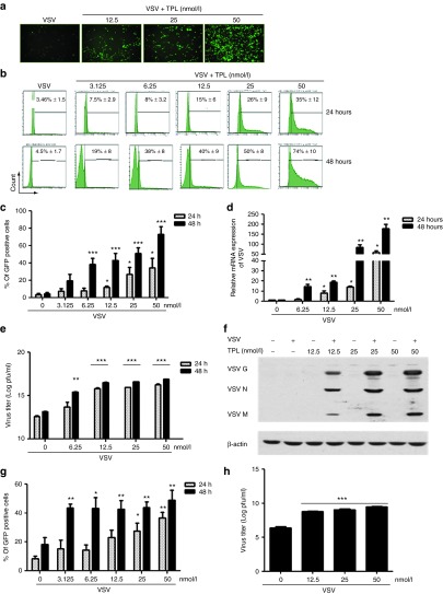
Triptolide (TPL) enhances vesicular stomatitis virus (VSV) viral replication in PC3 and other resistant cell lines in a dose- and time-dependent manner. PC3 cell line was either pretreated or not with indicated doses of TPL for 30 minutes (TPL was present in the medium). Cells were infected with VSV-Δ51-GFP (0.005 MOI) for 1 hour. The images were captured at 48 hours postinfection by fluorescent microscopy (Axiovert 40 CFL, Zeiss, Thornwood, NY) at a (a) original magnification ×10. Then, 24 and 48 hours postinfection, percentage of infected cells was determined by (b and c) flow cytometry analysis of GFP expression. VSV mRNA expression was examined by quantitative real-time PCR and the VSV mRNA level is displayed (d) as fold expression relative to the untreated VSV-infected sample. Virus released from infected cells was measured by (e) plaque assay and (f) western blotting for measuring viral proteins using anti-VSV antibody was also performed. DU145 and Karpas-422 cell lines were pretreated as described above. Cells were infected with VSV-Δ51-GFP (0.005 MOI for DU145 and 1 MOI for Karpas-422) for 1 hour. Then, 24 and 48 hours postinfection, surface expression of (g) VSV on DU145 was assessed by flow cytometry. Virus released from Karpas-422 infected cells was measured by (h) plaque assay. Histograms shown are representative of five independent experiments, and the values represent the means ± SEM for three to five independent experiments. For all the bar graphs, *P ≤ 0.05; **P ≤ 0.01; ***P ≤ 0.001 when compared with untreated VSV-infected cells.

