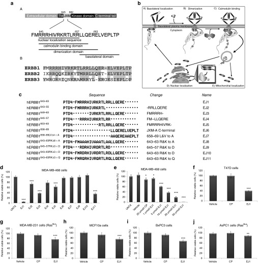
Figure 1.
Juxtamembrane domain peptides reduce cell viability. (a) (A) Schematic diagram depicting relevant functional motifs of the ERBB1 juxtamembrane (jxm) domain and (B) jxm domains of ERBB1, ERBB2, and ERBB3, with conserved regions based on National Center for Biotechnology Information protein alignment highlighted in gray. (b) Model of interactions involving the ERBB1 jxm. (A) ERBB1 localizes through its jxm (A')-contained targeting domain to the basolateral plasma membrane. (B) Ligand binding induces conformational changes in ERBB1, whereby interactions between jxm645–663 and the plasma membrane are disrupted, allowing dimerization and trans-phosphorylation. (C) Ca2+ influx promotes ERBB1 jxm domain interactions with proteins such as calcium-bound calmodulin (Ca2+/CaM) (C'). (D) Internalization and association with proteins such as importins α/β (D') and trafficking to locations such as the nucleus and mitochondria (D, E). (c) The amino acid number of ERBB1 is shown in the left column, corresponding to the specific amino acids shown in the middle column (sequence). Peptide designations are indicated in the right column. Changes from EJ1 in EJ2–11 are denoted in the second column from the right. (d–j) Cell lines were treated daily with 20 μmol/l EJ1, 20 μmol/l CP, or vehicle (water) for 3 days unless otherwise noted, and cell viability was determined by the MTT assay. Growth rates for vehicle-treated cells were set to 100%, and CP and EJ1 rates were adjusted accordingly. *P < 0.05, **P < 0.01, ***P < 0.001 (Student's t-test). Error bars, mean ± SD.

