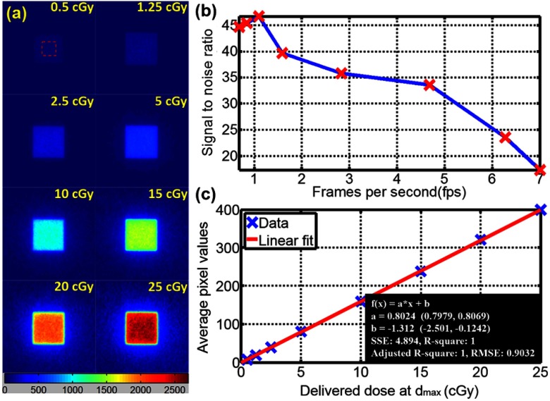
Fig. 2.
(a) Cherenkoscopy from a 6 MV square () photon beam incident normally on a tissue phantom () of opaque water equivalent phantom at with delivered dose at varying from 0.5 to 25 cGy. (b) Signal-to-noise ratio of Cherenkov images with fps from 0.68 to 7. (c) Linearity between intensity of Cherenkov emission from the surface and delivered dose is established under these controlled conditions.

