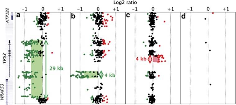
Figure 1.
Detection in LFS patients of CNVs targeting the TP53 locus, using a custom-designed 180K array CGH. Detection in three LFS patients, using custom-designed 180K array CGH of a complete deletion (a), a partial deletion removing the promoter region and exon 1 (b) and a duplication of exons 2–4 (c). (d) Probe coverage of the TP53 locus in the human catalogue 180K array CGH. CNVs detected by the ADM-2 algorithm are indicated by shaded area (green for deletion; red for duplication). Sizes of rearrangements are represented by double-headed arrows. Mapping of the corresponding genomic regions with the name of the genes (ATP1B2, TP53, WRAP53) are indicated on left.

