FIGURE 2.
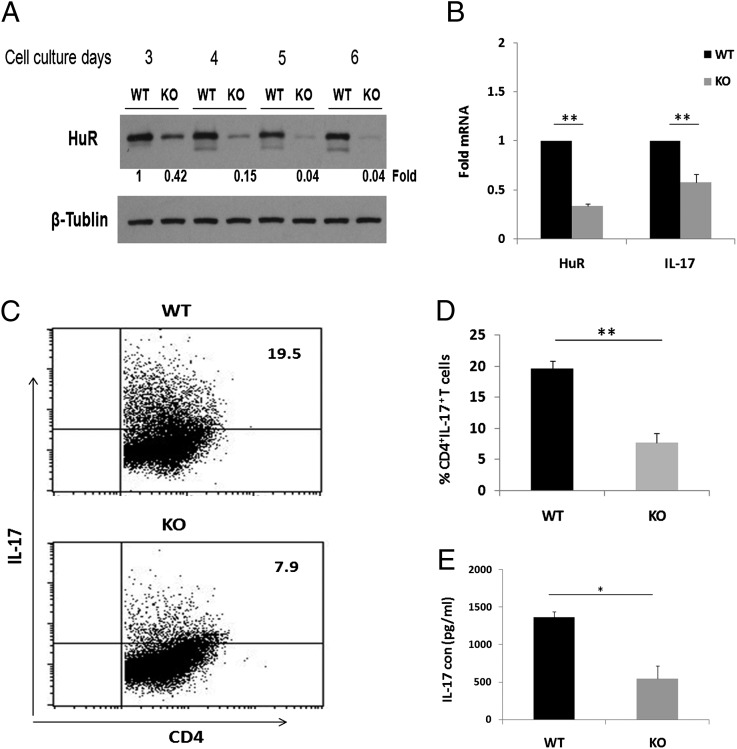
Reduced IL-17 expression in HuR KO Th17 cells. CD4+ T cells were isolated from spleens of control mice and HuR-deficient mice. Cells were stimulated as described in Fig. 1 (activation or Th17 polarization) for 3, 4, and 5 d. (A) HuR protein levels in Th17 cells were detected by Western blot analysis. HuR protein levels began to diminish starting on day 3 and were hardly detected on days 5 and 6 in KO Th17 cells. (B) Cells were collected at day 4. RNA was extracted, and quantitative RT-PCR was performed following reverse transcription using primer sequences listed in Table I. Decreased IL-17 mRNA was associated with reduced HuR mRNA in KO Th17 cells. IFN-γ mRNA levels were unaffected by HuR knockdown (data not shown). (C) CD4+ T cells were isolated from spleens of control mice and HuR-deficient mice. Cells were stimulated with plate-coated anti-CD3 (10 μg/ml) plus anti-CD28 (3 μg/ml) (activation) or Th17 cell–polarizing cytokines (Th17 polarization) as described in Materials and Methods. Cells were collected at day 5, and intercellular staining was performed for flow cytometric analysis. (D) HuR KO Th17 cells produced lower percentage of IL-17+ than WT Th17 cells. (E) Cell supernatant was collected at day 4, and IL-17 cytokine levels were measured by ELISA. KO Th17 cells produced lower levels of IL-17 protein than WT Th17 cells. Error bars represent ± SEM. Data were derived from at least three independent experiments. *p < 0.005, **p < 0.001.

