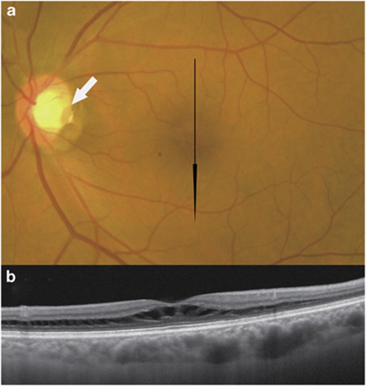
Figure 1.
Fundus photograph and swept-source optical coherence tomographic (SS-OCT) image of the left eye of a 66-year-old man with an optic disc pit. The light source of this SS-OCT system is a wavelength tunable laser centred at 1050 nm. (a) Colour fundus photograph. A grey, oval-shaped optic disc pit (white arrow) at the temporal margin of the disc and peripapillary pigmentary changes are observed. The optic disc tissue other than the pit seems normal. Glaucomatous cupping is not seen. Black arrow indicates the direction of the OCT scan. (b) Vertical SS-OCT image through the fovea showing retinoschisis. Tissue columns connecting the schisis cavities can be seen.

