
Figure 3.
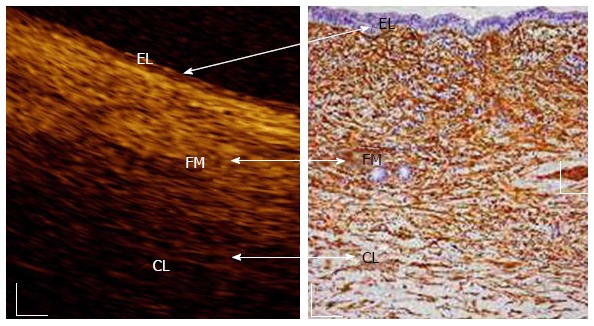
In vivo optical coherence tomography image of a normal main pancreatic duct wall compared with histology. Three recognizable layers were observed from the surface of the duct to a depth of 1 mm (Color online). The inner single layer of epithelial (EL) cells (400-800 μm thick) is visible as a superficial, hypo-reflective layer. The intermediate, connective fibro-muscular (FM) layer surrounding the epithelium, is visible as a hyper-reflective layer (350-600 μm thick). The outer connective-acinar (CL) structure close to the ductal wall epithelium is visible as a hypo-reflective layer[58]. White scale bar: 150 μm (right image).