
Figure 5.
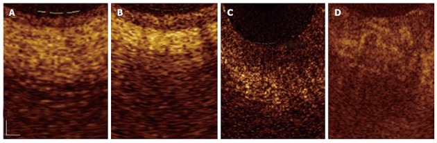
Magnified optical coherence tomography images. A: Sections with normal main pancreatic duct (MPD) wall; B: The presence of chronic pancreatitis; C: Low-grade dysplasia; D: Adenocarcinoma. Three differentiated layer architecture with a linear, regular surface, and a homogeneous back-scattered signal from each of the layer was observed in the normal condition. In the presence of chronic pancreatitis the optical coherence tomography (OCT) image still showed three-layer architecture, however, the inner epithelial layer appeared slightly larger than normal and the intermediate layer appeared more hyper-reflective; probably due to the presence of the dense mononuclear cell infiltrate. In the presence of dysplasia, OCT image showed thickened, strongly hypo-reflective and hetero-geneous inner MPD layer. Irregular surfaces were observed in the whole MPD structure. None of these layers were recognizable in the presence of adenocarcinoma[66]. Scale bar: 200 μm (Color online).