
Figure 8.
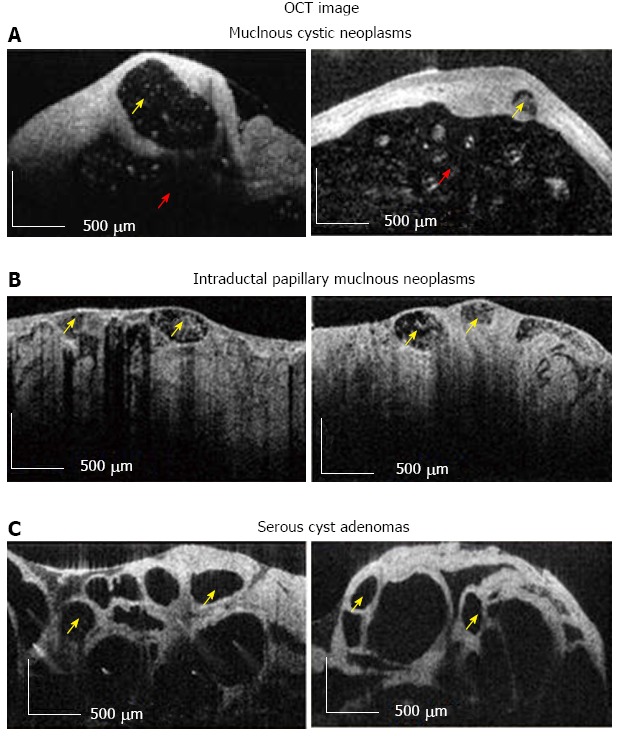
Optical coherence tomography image. A, B: Diagnostic criteria for high-risk (i.e., Mucinous Cystic Neoplasms, Intraductal Papillary Mucinous Neoplasms); C: Low risk (i.e., Serous Cysts Adenomas) pancreatic cysts. Multiple small cysts are marked with yellow arrow, while surrounded main cystic cavity is marked with red arrow. Scale bar = 500 μm[69]. OCT: Optical coherence tomography.