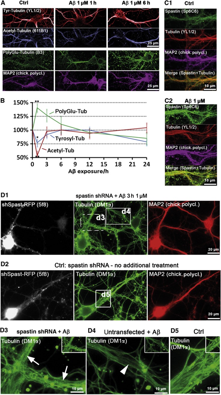
Figure 2.
Aβ exposure leads to rapid loss of acetylated, but not tyrosinated MTs, induces MT polyglutamylation and spastin recruitment in dendrites. Primary rat hippocampal neurons (16–21DIV) were treated with 1 μM Aβ as indicated, MAP2 staining was used as a marker for somatodendritic compartment. (A) In control cells, dendrites display high levels of tyrosinated and acetylated MTs, and low levels of polyglutamylated MTs. After short treatments (1 and 3 h), there is almost no change in tyrosinated MTs (upper panels), but a fast drop in acetylated MTs (middle panels). Polyglutamylation of MTs increases already after 1 h. Changes in all MT modifications are not observable anymore after 6 h of treatment (right panels). (B) Quantification of (A); changes revert to baseline after 6–12 h of treatment. (C) Cells were co-stained for spastin and MTs. (C1) Controls show only a diffuse spastin staining. (C2) After treatment with Aβ (1 μM, 3 h), spastin is recruited to MTs (increased colocalization with Tubulin in merge, lower panel). (D) Spastin was silenced using shRNA with a vector co-expressing RFP (5 days), cells were then treated with 1 μM Aβ for 3 h (D1, D3, D4) or left untreated (D2, D5). (D1) Silencing of spastin results in stable MTs after Aβ treatment. Cells expressing shRNA (RFP-positive cells, dotted box, magnified in D3, arrows) show no MT reduction as neighbouring untransfected cells do (RFP-negative cells, solid box, magnified in D4, arrowheads). (D2) Silencing of spastin in control cells has no effect on microtubule density. (D3–D5) Magnification of boxed areas in (D1) and (D2).

