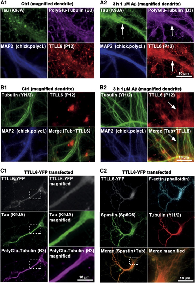
Figure 3.
TTLL6 induces polyglutamylation of MTs and Tau missorting. (A, B) Primary rat hippocampal neurons 21DIV treated with 1 μM Aβ for 3 h. MAP2 was used as a marker for the somatodendritic compartment. (A) Increases in dendritic polyglutamylation of MTs correlate with dendritic appearance of TTLL6 and Tau missorting (note colocalization indicated by arrows in A2) after Aβ treatment. Only basal/background levels of polyglutamylation of MTs, and no presence of TTLL6 or Tau can be detected in controls (A1). (B) Fixation and extraction to preserve MTs indicate recruitment of TTLL6 to MTs after Aβ treatment (B2; arrows), no recruitment is apparent in the case of controls (B1). (C) YFP-tagged TTLL6 was transfected into primary neurons for 1 day. (C1) Transfection of TTLL6 results in increased polyglutamylation and missorting of Tau. Right panels show magnification of boxed areas. (C2) TTLL6-transfected cells show enhanced spastin recruitment to MTs; merged images of spastin and tubulin show colocalization (lower right panel shows magnification of boxed area in lower left panel).

