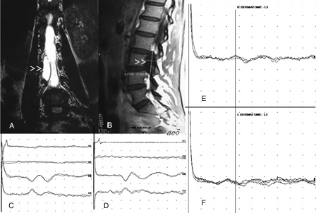
Figure 1.

Coronal T2 W (A) and sagittal T1W (B) MR images show a cystic lesion on the right side of the spinal canal (arrowheads). Tibial SEPs are normal bilaterally (C and D, Trace 1: peripheral, Trace 2: spinal, Traces 3 and 4: cortical responses). In dSEP studies, cortical responses to right L3 stimulation (E) has prolonged latency in comparison to that recorded with contralateral stimulation (F).
