
Figure 2.
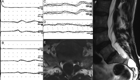
Tibial SEPs were normal bilaterally (A and B; the order of the traces are the same as in Fig. 1). The cortical response to right S3 dermatomal stimulation was normal (C) but was absent contralaterally (D). In the MR examination, very little spinal canal was left at the sacral level because of the huge arachnoid cysts in this patient. Marked scalloping is noted at the posterior margin of S2 vertebra (E and F).