
Figure 3.
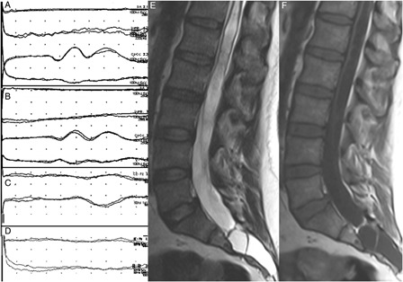
Tibial SEPs were normal bilaterally (A and B; the order of the traces are the same as in Fig. 1). The cortical response in right T11 dSEP study was normal (C) while it was not recordable contralaterally (D). Sagittal MR images show a cerebrospinal fluid containing-cystic lesion completely filling the sacral canal. Conus termination is at the L4 disc level. Distal cord is very thin due to the syringomyelic cavitation within it. Marked scalloping is noted in the posterior margin of the sacrum (E and F).