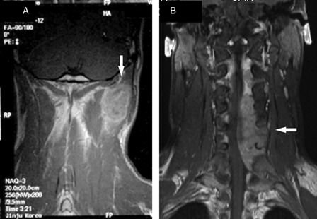
Figure 1.

Coronal T1-weighted post-contrast magnetic resonance images of the cervical spine. Magnetic resonance imaging scan of the cervical spine showing a large enhancing mass (arrow) surrounded by the trapezius, splenius, and levator scapulae muscles (A). Magnetic resonance imaging scan of the cervical spine obtained 1 year later showing a large extradural enhancing lesion (arrow), which is displacing the spinal cord to the right (B).
