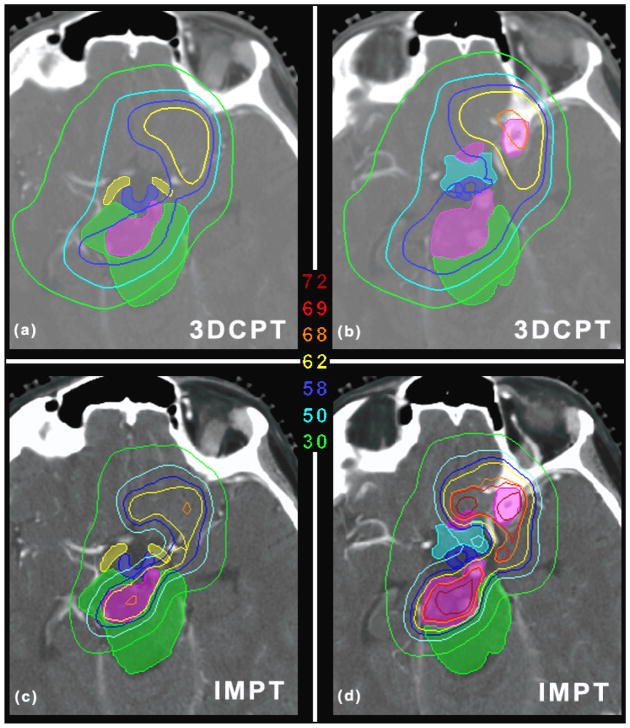
Figure 8.
(a) and (b) show the dose distribution for the clinical plan for two different axial slices for patient 2 (skull-base case). (c) and (d) are the same as (a) and (b) but for the IMPT plan. The unit of the isolines is Gy (RBE). The shaded regions identify the GTV (pink) and the OARs, i.e., the brainstem (green), hypothalamus (blue), chiasm (cyan), optical tracks (yellow). Axial slice thickness of 1.25 mm.

