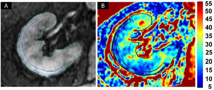
Figure 1.
Blood Oxygen Level Dependent (BOLD) MR: Selection of regions of interest (ROI) on an axial image: (A) Fractional tissue hypoxia was determined by outlining the entire axial kidney slice located within parenchyma. An additional ROI was placed to outline a “wide segment” cortical area excluding the renal collecting system, incidental cysts and the hilar vessels. (B) R2* parametric map for the selected axial slice reflecting widely variable R2* levels and deoxyhemoglobin at different sites within the kidney, particularly in medullary zones. This method of BOLD analysis bypasses observer selection of specific ROIs in the medulla and allows estimation of the fraction of the entire slice that exceeds the threshold above 30 sec-1 (see text).

