Fig. 2.

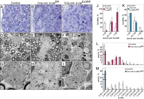
Impaired Wnt signaling in SCs causes defects in the sorting of axons and delays myelination. (A–C) Transverse semithin sections and (D–I) electron micrographs of peripheral nerves of control, Cnp-cre; β-catfl/fl, and Cnp-cre; β-catEx3fl/fl mice at P3. Note the large size of the axonal bundles in the Cnp-cre; β-catfl/fl nerves and the relatively small size of bundles in the Cnp-cre; β-catEx3fl/fl nerves. Sorted axons are marked with arrowheads, axonal bundles are marked with brackets, and unsorted large axons within the bundles are marked with arrows. (Scale bars: 10 μm in A–C and 5 μm in D–I.) Quantification of the axonal bundle size in control mice along with (J) Cnp-cre; β-catfl/fl (red) and (K) Cnp-cre; β-catEx3fl/fl (blue) mice. Shown is the percentage of bundles that contain a particular range of numbers of axons. Quantification of the g ratio in control mice along with (L) Cnp-cre; β-catfl/fl (red) and (M) Cnp-cre; β-catEx3fl/fl (blue) mice. Shown is the percentage of axons that display a particular g ratio. Errors bars indicate SD calculated from at least three mice per group.
