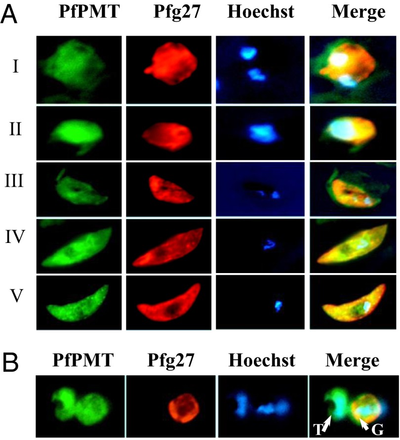
Fig. 1.
PfPMT expression during gametocyte development. (A) Wild-type 3D7 parasites were precultured at 2% parasitemia and 6% hematocrit in complete medium and maintained at 37 °C until the cultures reached 10% parasitemia. Parasites were then transferred to GM culture conditions and samples collected over time. Expression of PfPMT and the gametocyte-specific protein Pfg27 in gametocyte stages I, II, III, IV and V was monitored by immunofluoresence analysis using antibodies directed to PfPMT (green) or Pfg27 (red). Areas of overlap between PfPMT and Pfg27 appear in yellow. Nuclear staining was achieved using Hoechst 33258 (blue). (B) Culture sample showing adjacent trophozoite (T)- and gametocyte (G)-infected erythrocytes stained with anti-PfPMT and anti-Pfg27 antibodies.

