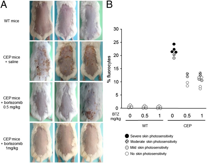
Fig. 4.
Skin photosensitivity reversion in CEP mice treated with bortezomib. (A) Mice were depilated and exposed to UVA irradiation (8 J/cm2). Representative macroscopic pictures of dorsal skin at 5 d after UVA irradiation are shown. (B) Porphyrin-accumulating RBCs (fluorocytes) were analyzed by flow cytometry in WT and CEP mice treated with 0.5 or 1 mg/kg bortezomib (BTZ). Each individual mouse is represented by a circle according to the macroscopic severity of skin photosensitivity: clear-open to dark-filled circles indicate the intensity of cutaneous lesions.

