
Fig. 6.
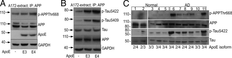
ApoE4 triggers APP-Thr668 phosphorylation and Tau phosphorylation. (A and B) Twenty-four hours after transfecting A172 cells with ApoE4, cell extracts were subjected to IP with anti-APP antibody followed by SDS/PAGE and WB to detect p-APP (A), p-TauSer422 (B, Top), p-TauSer409 (B, Middle), or Tau (B, Bottom). The last two panels in A represent ApoE and GAPDH (loading control) or APP and GAPDH (loading control) before the pull-down (B). (C) p-APP and p-Tau in AD. A total of 100 μg each of temporoparietal extracts isolated from normal and AD brains (Table S2) were examined by WB using APP, p-APPThr668, Tau, and p-TauSer422 antibodies. Isoform genotyping of ApoE was performed as described in SI Materials and Methods (Fig. S5).