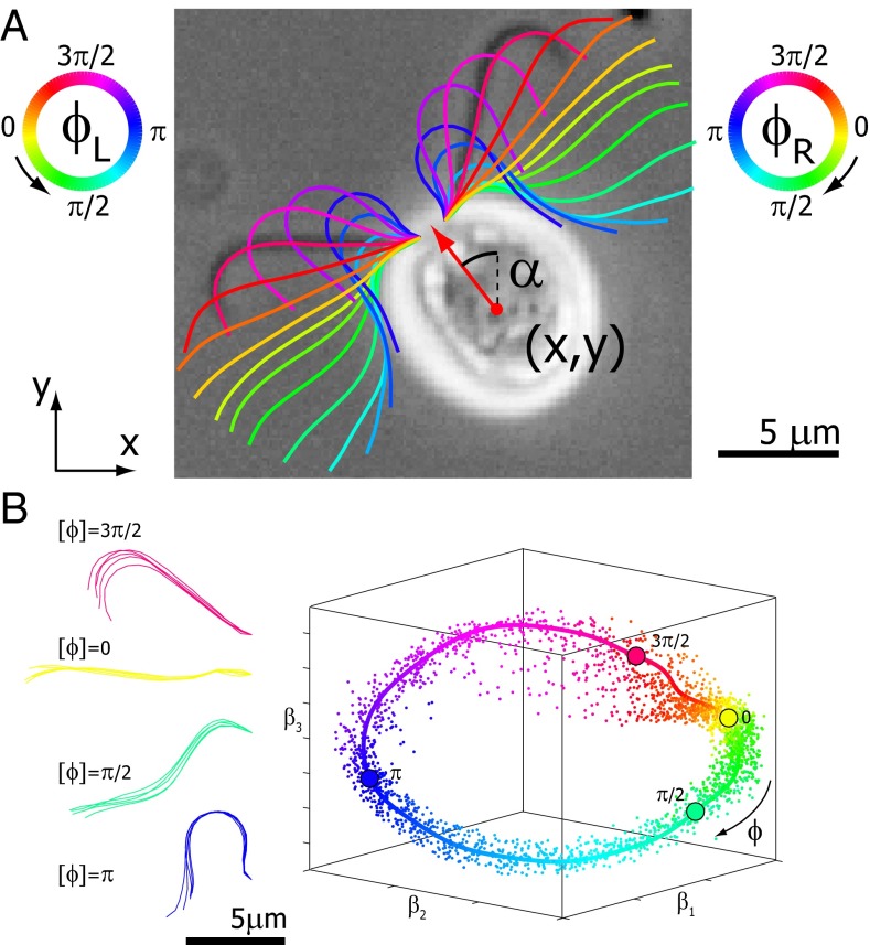
Fig. 1.
Five degrees of freedom for Chlamydomonas. (A) In our experiments, conducted in shallow observation chambers, Chlamydomonas cells swim in a plane. At each time, the position and orientation of the cell body is characterized by its center position  and the angle α of its long axis with respect to the laboratory frame. The beating of each flagellum is characterized by a single periodic phase variable,
  and
and  for the left and right flagellum, respectively. The flagellar shapes shown in different colors were tracked from high-speed recordings and correspond to a time-difference of
  . This beat pattern was used for all computations. (B) Binning of tracked flagellar shapes according to shape similarity defines a flagellar phase angle as shown on the left. More precisely, we employed a nonlinear dimensionality reduction technique as specified in Supporting Information to represent each tracked planar flagellar shape as a point in an abstract shape space. This representation reveals the periodicity of the flagellar beat and supports our description of the flagellar beat as a fixed sequence of flagellar shapes parameterized by a single phase variable φ.

