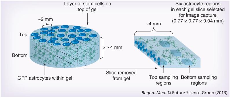
Figure 1. 3D cell culture system.
Astrocyte gels were set within 24-well culture plates before control or test cells were seeded onto the top surface. Following 5 days in culture, gels were fixed and a slice approximately 2-mm thick was removed from the center of each gel for confocal microscopy. Images were captured using standardized microscope settings from three positions at the top and three positions near the bottom of each gel slice.

