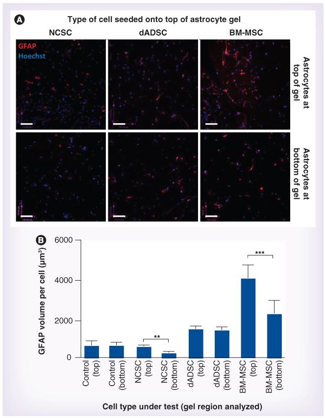
Figure 3. Astrocyte reactivity in response to test cell populations.
(A) Astrocytic GFAP immunoreactivity (red) was detected in defined locations within gels after 5 days in response to NCSCs, dADSCs or BM-MSCs seeded onto the top of gels. Astrocytes within the gels were distinguished from cells seeded on the surface using GFP, and nuclei were stained using Hoechst (blue). Scale bars = 100 μm. (B) Confocal image series were analyzed to quantify the volume of GFAP immunoreactivity per astrocyte within the gel regions. Three replicate series were analyzed within each region (top and bottom) of each gel and data are means ± standard error of the mean from at least six independent gels. Top and bottom regions were compared using a paired t-test. **p < 0.01; ***p < 0.001. BM-MSC: Mesenchymal stem cell from bone marrow; dADSC: Differentiated Schwann cell-like adipose-derived stem cell; NCSC: Neural crest stem cell.

