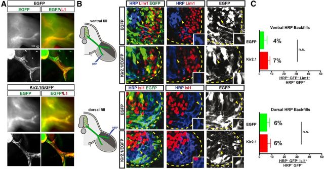
Figure 2.
Suppression of activity by Kir2.1 expression does not affect the fidelity of LMC motor axon limb trajectories. A, Detection of L1 and GFP proteins in wholemounted limbs (top) or their sections (bottom) in chick HH st. 17/18 embryos electroporated with EGFP or Kir2.1/EGFP at HH st. 27/28. L1 protein (red) labels peripheral nerves, while green fluorescence labels only GFP+ axons. No differences in outgrowth timing or pattern were noted. B, Retrograde labeling of LMC neurons by HRP injections into ventral or dorsal hindlimb shank muscles of chick HH st. 29/30 embryos expressing EGFP or Kir2.1/EGFP. Images show detection of HRP (blue), Lim1 or Isl1 (red), and EGFP (green) in the LMC region of EGFP- or Kir2.1/EGFP-electroporated embryos injected with HRP into ventral (top) or dorsal (bottom) shank muscles. Insets in images show Lim1− or Isl1− MN electroporated with EGFP or Kir2.1/EGFP and backfilled, as expected, by ventral or dorsal fills (indicated with yellow arrowheads). C, Proportions (%) of electroporated and backfilled lateral or medial LMC MN in embryos injected with HRP into ventral or dorsal shank muscles. In ventrally filled EGFP-expressing embryos, 4 ± 3% of HRP+, EGFP+ LMC neurons were Lim1+. Similarly, in ventrally filled Kir2.1/EGFP-expressing embryos, 7 ± 2% of HRP+, EGFP+ LMC neurons were Lim1+ (N = 4 for both EGFP+ and Kir2.1/EGFP+ embryos, n = 187 and 221 neurons, respectively). In dorsally filled EGFP-expressing embryos, 6 ± 2% of HRP+, EGFP+ LMC neurons were Isl1+. Similarly, in dorsally filled Kir2.1/EGFP-expressing embryos, 6 ± 3% of HRP+, EGFP+ LMC neurons were Isl1+ (N = 3 for both EGFP+ and Kir2.1/EGFP+ embryos, n = 172 and 293 neurons, respectively). Proportions of HRP+, EGFP+ lateral or medial LMC neurons in ventrally or dorsally filled Kir2.1/EGFP-expressing embryos and those in ventrally or dorsally filled EGFP-expressing embryos are not significantly different (p > 0.05; Student's t test). Error bars indicate SEM. All values are expressed and plotted as the mean ± SEM. Scale bars, 40 μm.

