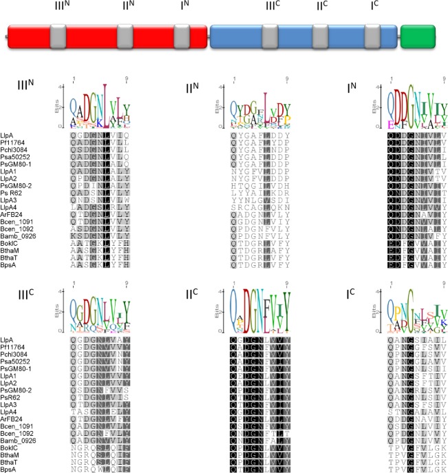
Figure 2.
General domain structure of LlpA proteins with potential carbohydrate-binding motifs (gray). The N-domain is colored red, the C-domain blue, and the C-terminal extension green. The respective potential mannose-binding motifs corresponding to the consensus motif QxDxNxVxY in LlpA-like proteins (derived from sequence alignment in Fig. S1) are aligned. The protein codes and accession numbers are specified in Figure 1. Sequence conservation is visualized by differential shading and the sequence logo graph visualizes the degree of consensus for each residue.

