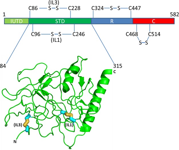
Figure 1.

Schematic representation of the domain arrangement of colE9 showing the positions of the disulfide bonds that have been generated in the STD, R, and DNase domains (see Table 1) and the crystal structure of colE3 from residues 84–315 (Soelaiman et al. 2001) that depict the STD with the locations of the corresponding cysteine mutations and consequent disulfide bond formation in the STD of colE9 for IL1 and IL3.
