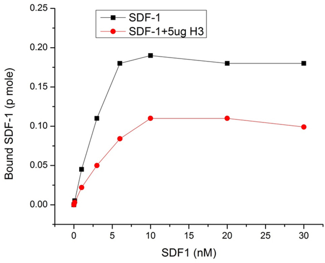
Figure 2. Effects of hBD-3 on saturatable SDF-1α binding to CEM cells.
The SDF-1α binding assay was performed as described in the Materials and Methods. 125I labeled SDF-1α (10nM) was added to each tube, together with 0 to 30 nM unlabeled SDF-1α in the absence or presence of 5µg/ml hBD-3. Cells were incubated at 37°C for 20min prior to termination of binding by rapid sedimentation and washing.

