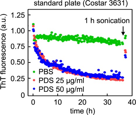
Figure 4.

PDS facilitates the adsorption of intact amyloid fibrils to plate well walls. Aβ1–40 fibrils were incubated in the absence (green) or in the presence of 25 or 50 µg/mL PDS (red and blue, respectively), as described in Figure 1(C). After 35 h of incubation, the plate was sonicated for 1 h, and the ThT fluorescence measured again. Most of the ThT fluorescence of PDS samples is recovered, indicating that Aβ fibrils are adsorbed to the well walls.
