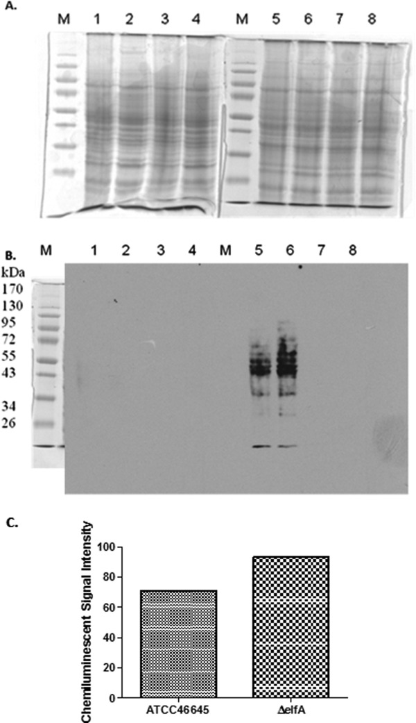
Figure 3.

Western blot analysis of protein glutathionylation in A. fumigatus ATCC46645 and ΔelfA. (A) SDS-PAGE of A. fumigatus ATCC46645 (Lanes 1, 3, 5, 7) and ΔelfA (Lanes 2, 4, 6, 8) whole cell lysates (50 µg) under reducing (Lanes 1–4) and nonreducing conditions (Lanes 5–8). The samples in Lanes 1, 2, 5, and 6 were incubated with biotin-GSSG, while the samples in Lanes 3, 4, 7, and 8 were incubated with water as a control. (B) Western blot probed with streptavidin-HRP. Proteins glutathionylated upon addition of biotin-GSSG were detected in A. fumigatus ATCC46645 (Lane 5) and ΔelfA (Lane 6). There were more glutathionylated proteins detected in A. fumigatus ΔelfA (Lane 6) than in A. fumigatus ATCC46645 (Lane 5). No glutathionylated proteins were detected on the reduced blot (Lanes 1–4) or in the samples treated with water (Lanes 7–8). (C) Image analysis indicates a 24% increase in chemiluminescent signal intensity in A. fumigatus ΔelfA compared to A. fumigatus ATCC46645 supporting the observation that there was a higher level of protein glutathionylation in A. fumigatus ΔelfA.
