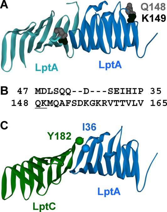
Figure 1.

Structures of LptA and LptC. (A) The LptA dimer observed in the 2R19 crystal structure12 with sites Q148 and K149 highlighted. The K149 side chain was not resolved and is missing from the structure. (B) The N-terminal amino acids 35–47 and the C-terminal amino acids 148–165 of LptA are aligned based on the crystallographic oligomer. The gap occurs because the C-terminal loop in the fold of the β-strand is longer than in the N-terminus edge strand. Amino acids K149 and Q148 are underlined. (C) Docked model of the LptC (green;3MY2)-LptA (blue; 2R19) interaction based on the LptA dimer structure. Reporter sites Y182 LptC and I36 LptA used for the affinity studies are labeled.
