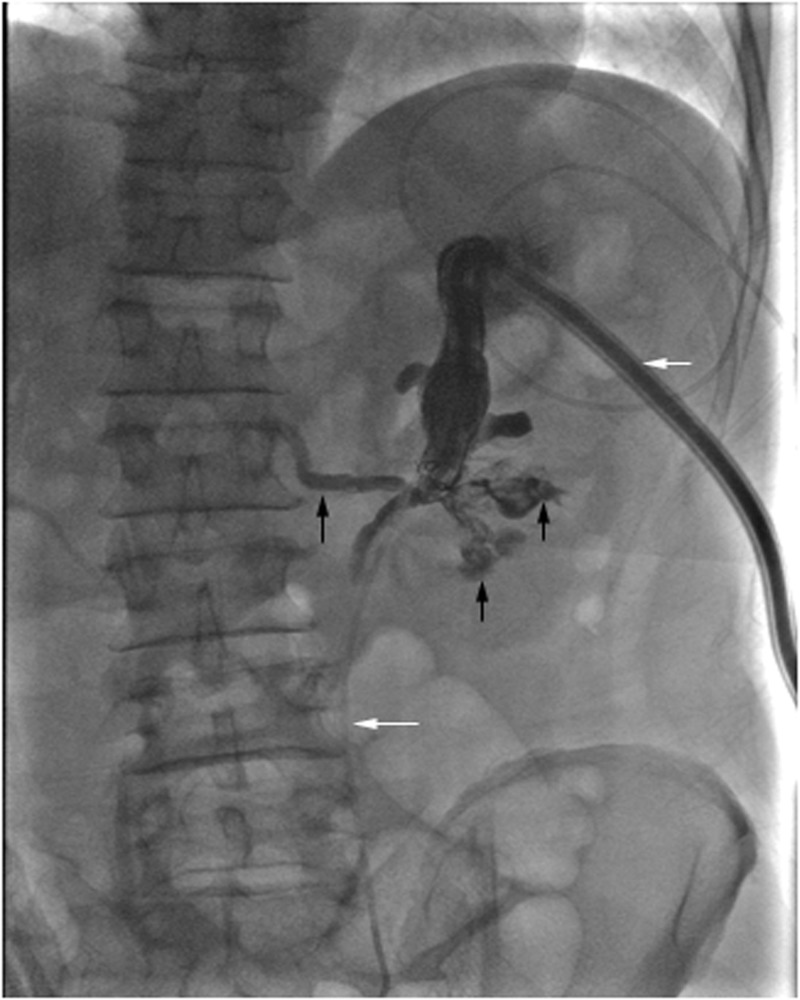
Fig. 3.
Nephrostogram following embolization of an AVF demonstrates nephrostomy catheter (short white arrow) and ureteral stent (long white arrow) positions in the upper pole calyx and ureter, respectively. Visualization of the residual clots in the renal pelvis and calyces (short black arrows), and immediate filling of posterior segmental vein (long black arrow), consistent with findings of coexisting caliceovenous fistula

