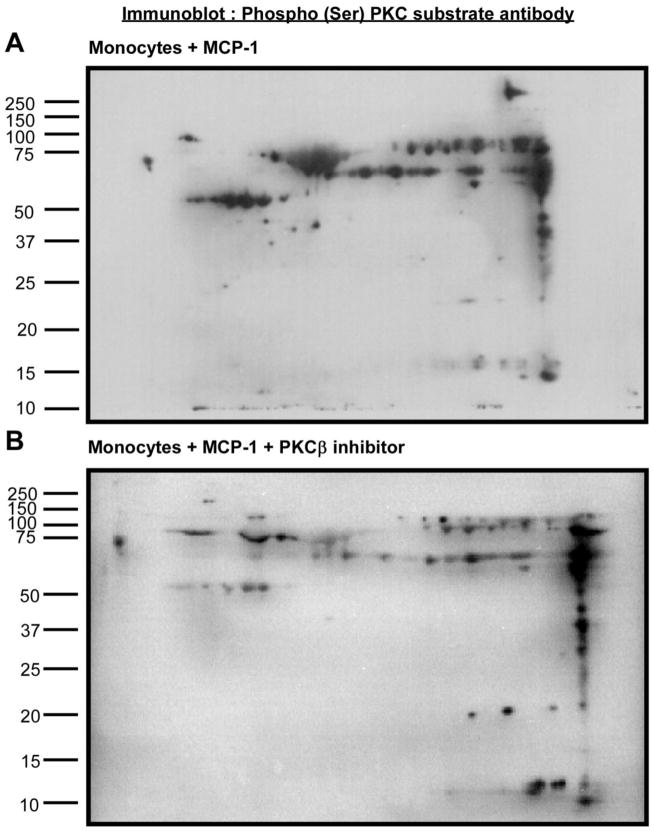
Figure 3. Detection of phosphorylated proteins in MCP-1-treated monocytes in the presence/ absence of PKCβ inhibitor peptide.
Human monocyte lysates were fractionated by 2-DIGE and immunoblots were probed with phosphor (Ser) PKC substrate antibody. Figure 3A shows the monocyte group treated with MCP-1 and Figure 3B shows the monocyte group treated with MCP-1+PKCβ inhibitor peptide.

