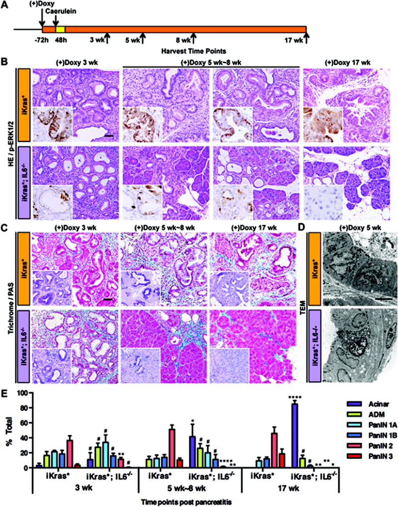
Figure 2. IL6 is necessary for PanIN progression and maintenance.

(A) Experimental design, n=4~7. (B) H&E and p-ERK1/2 staining (insets) of iKras* and iKras*;IL6−/− mice pancreata 3 weeks, 5 weeks~8 weeks and 17 weeks following pancreatitis. Scale bar 50μm. (C) Gomori Trichrome staining and Periodic Acid Schiff staining (insets) of iKras* and iKras*;IL6−/− mice pancreata. Scale bar 50μm. (D) Transmission electron microscope image of iKras* and iKras*;IL6−/− mice pancreata 5 weeks following pancreatitis. Scale bar 10μm. (E) Pathological analysis. Data represent mean ± SEM, n=3~5. The statistical difference between iKras* and iKras*;IL6−/− mice at the same time point per lesion type was determined by two-sided Student’s t-test. *p<0.05, **p<0.01, ****p<0.0001, #not significant.
